Call-an-Ambulance
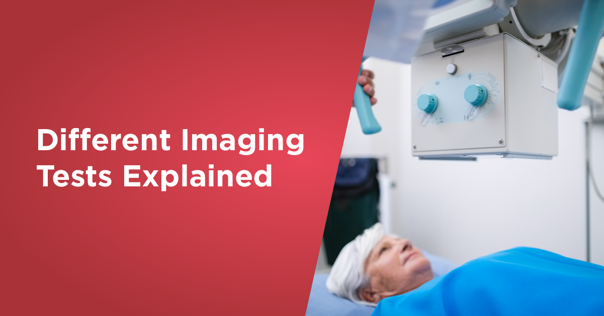
What is imaging test?
Imaging test is a process that provides comprehensive images of body parts on a macroscopic level. Imaging tests employ a variety of energy sources like radio waves, radioactive substances, ultrasound, and X-rays. Image processing is used to understand the detailed condition of an ailment, treatment planning, or monitor the efficacy of treatment. Computed tomography (CT), mammography, ultrasonography, magnetic resonance imaging (MRI), and nuclear medicine exams are some popular types of imaging tests.
Many imaging procedures are simple and painless. Diagnostic imaging involves spending a lot of time motionless within a machine. Some of these processes can lead to light exposure to radiation.
The five most common radiological tests prescribed by doctors are explained below.
CT Scan
CT scan uses a series of X-rays to create crosssections of the inside of the body, including bones, blood vessels, and soft tissues.
What to Expect: You will lie on a table that slides into the scanner, which looks like a large doughnut. The X-ray tube rotates around you to take images.
Imaging Method: Ionizing Radiation
Duration: 10-15 minutes
Used to Diagnose:
- Bone fractures
- Tumours and cancers
- Vascular disease
- Heart disease
- Infections
- Used to guide biopsies
- Aneurysms
- Injuries from trauma
MRI
MRIs use magnetic fields and radio waves to create detailed images of organs and tissues in the body.
What to Expect: You lie on a table that slides into the MRI machine, which is deeper and narrower than a CT scanner.
Imaging method: Magnetic Waves
Duration: 45 minutes – 1 hour
Used to Diagnose:
- Brain Disease
- Multiple Sclerosis (MS)
- Stroke
- Spinal cord disorders
- Tumours
- Blood vessel issues
- Joint or tendon injuries
Ultrasound
Ultrasound uses high-frequency sound waves to produce images of organs and structures within the body.
What to Expect: A technician applies gel to your skin, then presses a small probe against it, moving it to capture images of the inside of your body.
Imaging Method: Sound Waves
Duration: 15 – 25 min
Used to Diagnose:
- Gallbladder disease
- Breast lumps
- Genital/prostate issues
- Joint inflammation
- Blood flow problems
- Monitoring pregnancy
- Used to guide biopsies
PET Scan
PET scans use radioactive drugs (called tracers) and a scanning machine to show how your tissues and organs are functioning.
What to Expect: You swallow or have a radiotracer injected. You then enter a PET scanner (which looks like a CT scanner) that reads the radiation given off by the radiotracer.
Imaging Method: Radiotracers
Duration: 1.5-2 hours
Used to Diagnose:
- Cancer
- Heart disease
- Coronary artery disease
- Alzheimer’s Disease
- Seizures
- Epilepsy
- Parkinson’s Disease
X-Ray
X-rays are quick, painless tests that produce images of the structures inside your body especially bones
What to Expect: you will lie, sit, or stand while the X-ray machine takes images. You may be asked to move into several positions.
Imaging Method: Ionizing Radiation
Duration: 10-15 minutes
Used to Diagnose:
- Bone fractures
- Arthritis
- Osteoporosis
- Infections
- Breast cancer
- Swallowed items
- Digestive tract problems
Advanced facilities and imaging tests are available at Regency Hospitals in Kanpur and Lucknow.
Request a call back